Dental disease is not merely a cosmetic concern in companion animals. It can cause pain, infection, tooth loss, and difficulty eating. The American Veterinary Dental College reports that periodontal disease affects more than 80% of dogs and 70% of cats by three years of age. These figures reveal a serious clinical need, not a minor grooming issue.
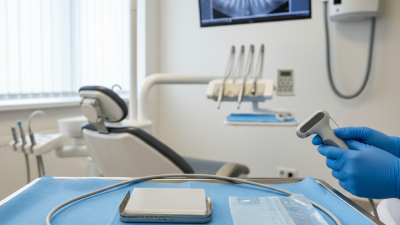
A veterinary dental x ray machine helps clinicians see what oral examination cannot reveal. It shows root fractures, retained roots, bone loss, and lesions beneath apparently normal crowns. The 2019 AAHA Dental Care Guidelines recommend full-mouth dental radiography because many important abnormalities remain hidden below the gumline. During a procedure, a small sensor can capture the root apex beside a molar within seconds. That image may change an extraction decision, anesthesia plan, or follow-up recommendation.
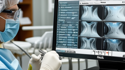
The equipment also supports clearer communication with pet owners. A visible radiograph often explains pain better than a verbal description. It can strengthen informed consent and create a reliable medical record. However, technology alone is not enough. Poor positioning, motion, or limited training can produce weak images. I have to admit this point is easy to underestimate. A costly machine may remain underused without workflow planning and staff practice.
Choosing the right system therefore requires more than comparing megapixels. Clinics should assess image quality, sensor durability, software integration, radiation safety, maintenance support, and staff training. Used responsibly, dental radiography improves diagnostic confidence and may help preserve healthy teeth. Its greatest value is practical: finding hidden disease before it becomes impossible to ignore.
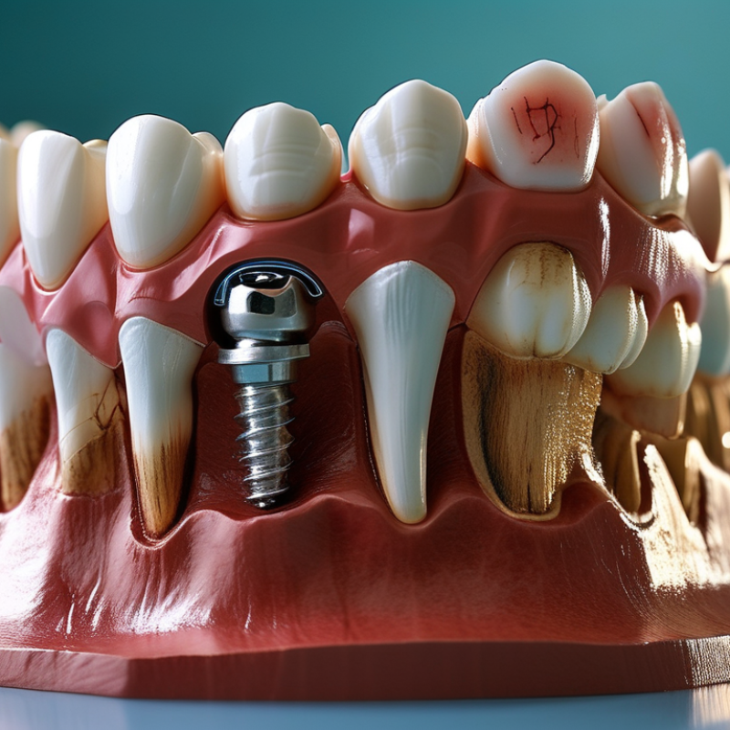
A veterinary dental X-ray machine is specialized imaging equipment for examining teeth, roots, and surrounding bone in animals. It usually uses an intraoral sensor or dental film to capture focused images inside the mouth. Unlike a routine oral inspection, dental radiography can show hidden root fractures, bone loss, retained teeth, and lesions beneath the gumline.
The need is substantial. The 2019 AAHA Dental Care Guidelines report periodontal disease in over 80% of dogs and 70% of cats by three years of age. These figures make visual examination alone feel insufficient. A clean-looking crown may hide a damaged root. Small details matter.
During a dental procedure, the machine helps veterinarians assess each tooth before extraction or treatment. Clear images can reduce guesswork, shorten repeated handling, and support more precise records. WSAVA dental guidance also emphasizes radiographic evaluation when disease extends below the gumline. However, image quality depends on positioning, exposure settings, sensor care, and staff training. Technology does not replace judgment. It can even create false confidence when technique is poor.
A useful system should provide sharp images, practical software, adjustable exposure control, and comfortable positioning for different animal sizes. Radiation safety remains essential, including controlled access and appropriate protective procedures. The best choice is not always the most advanced machine. It is the one that produces consistent diagnostic images in real clinical conditions.
| Data Dimension | Information | Why It Matters in Veterinary Dentistry |
|---|---|---|
| Definition | A veterinary dental X-ray machine is an intraoral radiography system designed to capture detailed images of teeth, roots, periodontal structures, and surrounding jawbone in animals. | It provides diagnostic information that cannot be obtained reliably through a visual oral examination alone. |
| Primary Application | Dental examination, periodontal assessment, extraction planning, oral surgery, endodontic evaluation, and post-treatment review. | The images help support treatment planning and reduce the risk of leaving diseased or fractured root material behind. |
| Image Area | The system is intended for small intraoral image receptors placed close to individual teeth or small groups of teeth. | The small field of view supports high-detail imaging in the compact oral anatomy of dogs, cats, and other small animals. |
| Common Tube Voltage | Many intraoral dental systems operate within an approximately 60–70 kVp range, although the exact specification depends on the equipment design. | Tube voltage affects X-ray penetration and image contrast. The selected setting should follow the equipment instructions and clinical protocol. |
| Exposure Control | Exposure is controlled through selectable time, tube-current, or combined exposure settings, depending on the system. | Accurate exposure control helps produce diagnostic images while avoiding unnecessary radiation exposure. |
| Image Receptor Options | Compatible receptors may include digital intraoral sensors, photostimulable phosphor plates, or dental film, depending on the workflow. | Digital receptors can speed up image review, while plate-based systems may offer flexibility in positioning and handling. |
| Typical Dental Findings | Periodontal bone loss, retained roots, tooth resorption, periapical disease, pulp exposure, fractured teeth, unerupted teeth, and jaw lesions. | Radiographs reveal structures below the gumline and inside the tooth that may appear normal during an external examination. |
| Patient Positioning | The animal is generally positioned according to the dental region being examined, with the receptor placed intraorally and the X-ray tube aligned with the target area. | Correct positioning reduces distortion and improves visualization of tooth roots and surrounding bone. |
| Anesthesia Requirement | Complete veterinary dental radiography commonly requires general anesthesia or an appropriate controlled anesthetic protocol. | Anesthesia helps prevent movement, allows accurate intraoral positioning, and supports a thorough oral examination and treatment. |
| Diagnostic Benefit | Provides two-dimensional radiographic views of dental and jaw structures that are not visible externally. | Veterinary professionals can make better-informed decisions about extraction, periodontal therapy, root canal treatment, or further investigation. |
| Workflow Advantage | A dedicated dental system is designed for frequent intraoral imaging and can be integrated into a veterinary dental treatment area. | A suitable setup can reduce repositioning time, improve image consistency, and support efficient dental procedures. |
| Radiation Protection | Safe operation requires appropriate shielding, controlled access, equipment maintenance, dosimetry practices where required, and compliance with local regulations. | Radiation safety protects veterinary staff and other people while maintaining a controlled imaging environment. |
| Maintenance Considerations | Routine checks should include tube-head condition, exposure controls, cables, receptor compatibility, image quality, and required regulatory inspections. | Regular maintenance supports reliable imaging, consistent diagnostic quality, and safer long-term operation. |
| Best-Suited Users | Small-animal hospitals, general veterinary practices performing dental procedures, referral dental services, and veterinary teaching facilities. | The equipment is especially useful where dental assessment and treatment are performed regularly. |
Note: Technical specifications, operating procedures, and legal requirements vary by equipment type and location. Always follow the manufacturer’s instructions and applicable radiation-safety regulations.