How to Choose a Handheld X-Ray Machine?

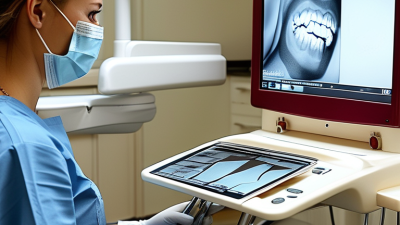
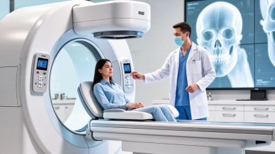
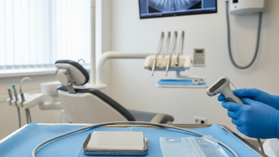
Choosing a hand held x ray machine is not just a matter of finding the smallest unit or the lowest price. The right choice depends on where it will be used, which examinations it must support, and who will operate it. A clinic examining small animals has different needs from a dental practice or a mobile imaging service. Details matter: the grip should feel stable, controls should be easy to read, and the display should remain clear under bright room lights. A few minutes of hands-on testing can reveal problems that a product sheet cannot.

Marie Curie, a pioneering radiation scientist, wrote, “Nothing in life is to be feared, it is only to be understood.” Her words offer a useful reminder: buyers should understand both the machine’s capabilities and its limitations. Compare image quality, battery life, weight, exposure settings, detector compatibility, service support, and operator training. Ask for clear specifications and a practical demonstration. If a claim sounds impressive but lacks measurable detail, pause and verify it. That extra step is easy to skip.

This guide explains how to assess those factors without treating one model as right for every setting. Consult qualified radiography professionals and medical physicists when evaluating technical performance and safe workflows. Consider the daily routine, too: who carries the unit, where it is stored, and how downtime is handled. A compact machine can still be awkward after a long shift. That matters. The best choice is the one that fits documented clinical needs, performs consistently, and comes with dependable training and support.
How to Choose a Handheld X-Ray Machine?

Identify the Imaging Task and Applicable Standard: IEC 60601-2-65 for Dental Units

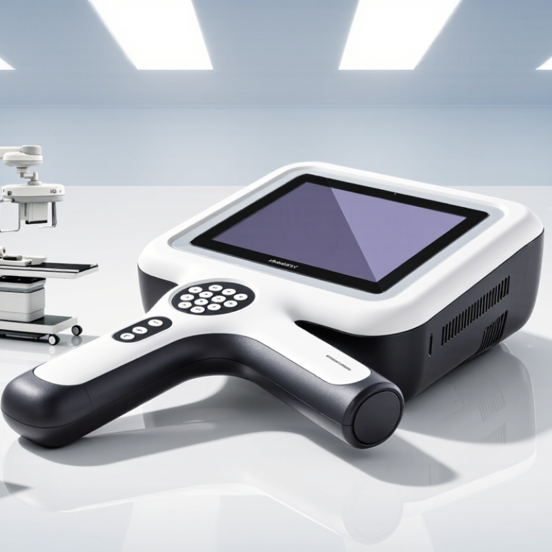
When choosing a handheld X-ray machine, define the imaging task before comparing features. Are you taking routine intraoral images, working in a small operatory, or needing mobile access between treatment rooms?

Match the unit’s output and exposure controls to the receptor, patient size, and required image detail. Check how easily the tube head aligns with a sensor; a few millimeters of drift can mean a retake.

Keep it practical.

IEC 60601-2-65 specifies particular basic-safety and essential-performance requirements for intraoral dental X-ray equipment. It should be considered alongside IEC 60601-1 and other applicable standards. Do not assume that every handheld design falls within the same scope.

Confirm the product’s intended use, classification, and conformity documentation with the manufacturer, and verify applicable requirements with a qualified testing or regulatory professional. Ask to review evidence for radiation output, exposure control, and essential performance—not just a general compliance statement.

Documentation matters.

Also assess practical protection: follow the unit’s instructions for positioning, operator distance, and any required shielding. A portable device can improve access, but it does not remove the need for trained users, controlled exposure, and a stable image receptor.

One detail is easy to overlook: a comfortable grip cannot compensate for poor positioning.
Compare Tube Voltage, Current, and Exposure Time for the Intended Exam

How to Choose a Handheld X-Ray Machine?

Compare Tube Voltage, Current, and Exposure Time for the Intended Exam

Start with the exam, not the highest available setting. Tube voltage, measured in kVp, affects beam penetration and image contrast. A thicker body part may need greater penetration, while a small extremity generally needs less. Tube current, measured in mA, controls how many X-rays the tube produces. Exposure time determines how long that output continues. Together, current and time form mAs, a useful measure of total tube output. They are related, but not interchangeable in every situation.

For a moving patient, a shorter exposure can reduce motion blur. Raising current may help maintain output during that brief exposure, if the machine supports it. For a still hand or foot, a longer exposure may be acceptable, but unnecessary output should still be avoided. Match settings to the body part, patient size, detector, and the manufacturer’s validated protocols. Small changes matter.

Use image feedback carefully. AAPM Task Group 116 describes a standardized exposure index calibration in which EI 100 corresponds to 1 μGy of detector air kerma under reference beam quality RQA5. This is detector exposure, not patient dose, and it is not a target for every exam. Review image quality and exposure indicators alongside routine quality-control results. A crisp image can still be overexposed. And protocols need checking.

For Dental Units, Verify the 0.5-mm Pb-Equivalent Backscatter Shield

How to Choose a Handheld X-Ray Machine?

For dental units, do not judge the backscatter shield by its appearance. Verify that documentation confirms at least 0.5 mm lead-equivalent protection, as specified for handheld X-ray equipment in ANSI/HPS N43.17-2009. Check that the shield is securely attached, undamaged, and positioned between the operator and the patient during exposure. A small gap or poor angle can matter. Ask for the test documentation, including the measurement conditions; “lead-equivalent” values are meaningful only when their testing basis is clear.

Shielding is one part of dose control, not a substitute for sound technique. ICRP Publication 103 recommends an occupational effective-dose limit of 20 mSv per year averaged over five years. This is a limit, not a personal target or proof that a particular unit is safe in every setup. Keep the shield close to the patient as directed by the manufacturer, use the unit’s prescribed hand position, and follow local radiation-safety guidance. I would not assume two shields perform alike just because both meet the same thickness figure.

Tips: Inspect the shield before use. Confirm its Pb-equivalent rating in writing, and arrange routine checks by a qualified radiation-safety professional. Record any damage or loose fittings; small defects are easy to overlook.

How to Choose a Handheld X-Ray Machine?

For dental units, verify the backscatter shield’s lead equivalence.

U.S. FDA criterion: hand-held X-ray equipment must have a protective barrier of at least 0.5 mm lead equivalent at 100 kVp (21 CFR §1020.30(h)(1)(i)). Check the device documentation for the shield’s stated lead equivalence and applicable test conditions; this chart is a reference criterion, not a measurement of any specific device.

Check Image Quality and Dose Against Acceptance-Test and ALARA Practices

How to Choose a Handheld X-Ray Machine?

Check Image Quality and Dose Against Acceptance-Test and ALARA Practices

Choose a handheld X-ray unit by reviewing its acceptance-test record, not just a sharp sample image. Ask a qualified medical physicist to measure tube voltage, exposure time, output consistency, beam alignment, and leakage. Use a suitable test phantom to assess contrast, detail, and image noise against the recorded baseline. Keep the geometry consistent. Small differences matter.

Dose checks need context. ICRP Publication 103 recommends an occupational effective-dose limit of 20 mSv per year, averaged over five years, with no single year above 50 mSv; the public limit is 1 mSv per year. These are protection limits, not targets for routine work or patient exposures. Apply ALARA by choosing exposure settings that produce diagnostically useful images without unnecessary dose, and follow applicable local requirements. A missed repeat image can matter, too.

Tips: Request the acceptance-test results before purchase. Confirm that the test includes dose-output measurements at the settings you expect to use. Check how the unit’s shield and positioning instructions reduce staff exposure, and arrange periodic quality-control checks. Record each result. If a test value drifts, investigate before relying on the device. I would not judge image quality from one ideal image alone.

How to Choose a Handheld X-Ray Machine? - Check Image Quality and Dose Against Acceptance-Test and ALARA Practices

Evaluation Area What to Check Acceptance-Test Approach ALARA and Selection Considerations
Image quality and diagnostic detail Assess sharpness, contrast, noise, and visibility of relevant anatomical details using the intended image receptor and a suitable dental test object or phantom. Record baseline images under documented exposure settings. Compare results with the receptor and system specifications, local acceptance criteria, and the clinic’s diagnostic requirements. Choose a system that produces clinically usable images without routinely needing higher exposure settings. Confirm compatibility with the clinic’s digital sensors or imaging plates.
Exposure settings and controls Review available kVp, mA, and exposure-time settings, along with patient-size selections and the clarity of control indicators. Verify displayed settings and operation according to the manufacturer’s instructions and applicable testing requirements. Document the settings used for baseline tests. Use the lowest exposure setting that consistently provides adequate diagnostic information for the patient and examination. Avoid relying on a single setting for every patient.
Exposure-time accuracy and reproducibility Check whether exposure duration is consistent across repeated exposures at the same selected setting. Measure with appropriate test equipment following the device instructions and local acceptance-test protocol. Compare results with applicable tolerances; do not assume one tolerance applies in every jurisdiction. Reliable exposure timing helps prevent unnecessarily long exposures and reduces the chance of repeat images caused by inconsistent output.
X-ray output and beam quality Assess output consistency and, where required, tube potential and beam quality using calibrated equipment. Have a qualified person perform the tests with suitable instruments. Compare measurements with the manufacturer’s specifications and relevant regulatory or professional requirements. Use measured baseline data to support technique selection and future quality-control comparisons rather than judging dose from image appearance alone.
Patient dose and dose monitoring Measure an appropriate dose indicator, such as air kerma or dose-area product where applicable, using a suitable dosimeter and a documented setup. Record the measurement conditions, exposure settings, receptor, and phantom or test geometry. Compare results with local diagnostic reference levels where available and investigate unexpected changes. There is no single dose value that is appropriate for every device, examination, patient, or jurisdiction. Optimize exposure for the individual examination and avoid unnecessary repeats.
Collimation and beam alignment Check beam size, centering, and alignment with the image receptor; confirm that any supplied positioning or collimation accessories fit securely. Evaluate the beam and receptor relationship using an appropriate test method. Confirm compliance with local requirements and the manufacturer’s specifications. Use the smallest practical field of view that covers the region of interest. Accurate positioning and alignment can reduce irradiation outside the area needed for diagnosis.
Backscatter shielding and operator protection Inspect the backscatter shield for correct installation, condition, and positioning according to the device instructions. Confirm the shield and other protective features are present and used as designed. Review the manufacturer’s operating instructions and the facility’s radiation-protection procedures. Follow local rules for operator location, orientation, and safe operating distance. Do not hold the device or expose patients in a manner that conflicts with regulations or the manufacturer’s instructions.
Workflow, training, and quality control Review ergonomics, weight, hand controls, battery status indicators, and whether the unit can be positioned consistently by trained users. Before clinical use, complete documented acceptance testing and user training. Establish routine checks, service records, and repeat testing after relevant repairs or performance concerns. Consistent positioning, exposure selection, and image review help limit retakes. Keep written protocols aligned with current local regulations and professional guidance.

Important: Acceptance-test limits, licensing rules, and radiation-protection requirements vary by location and equipment. Use the applicable regulations, manufacturer’s instructions, and a qualified radiation-safety professional to set and verify local criteria.

Assess Weight, Battery Capacity, and Workflow for Your Daily Exam Volume

A handheld X-ray unit should feel manageable after a full shift, not just during a showroom demonstration. Check its total working weight with the battery attached, then simulate reaching across a bed or holding the unit between repeated views. Small details matter. A heavy front end can make positioning slower and less steady. Ask how the device balances in your hand, and whether its grip remains comfortable with gloves. There is no universal “light enough” threshold; staff size, exam type, and positioning demands all matter.

Battery capacity must match real daily volume, including repeats, standby time, and travel between rooms. Do not rely on a headline exposure count alone. Record a typical busy day, then ask the supplier how many comparable exams one charge supports. WHO’s Global Tuberculosis Report 2024 estimated 10.8 million people developed TB worldwide in 2023, illustrating why mobile imaging workflows can face heavy demand in some settings. That figure is not a workload target for every clinic. Image quality still matters: WHO’s 2021 TB screening evidence review reported chest X-ray sensitivity of 94% and specificity of 89% when any abnormality counted as a positive screen. Build time for positioning and image review, not just exposures. I would still question optimistic battery estimates; local exam patterns can be messier than a test chart.